Conjunctival Melanosis

Conjunctival Melanosis

0 Comments

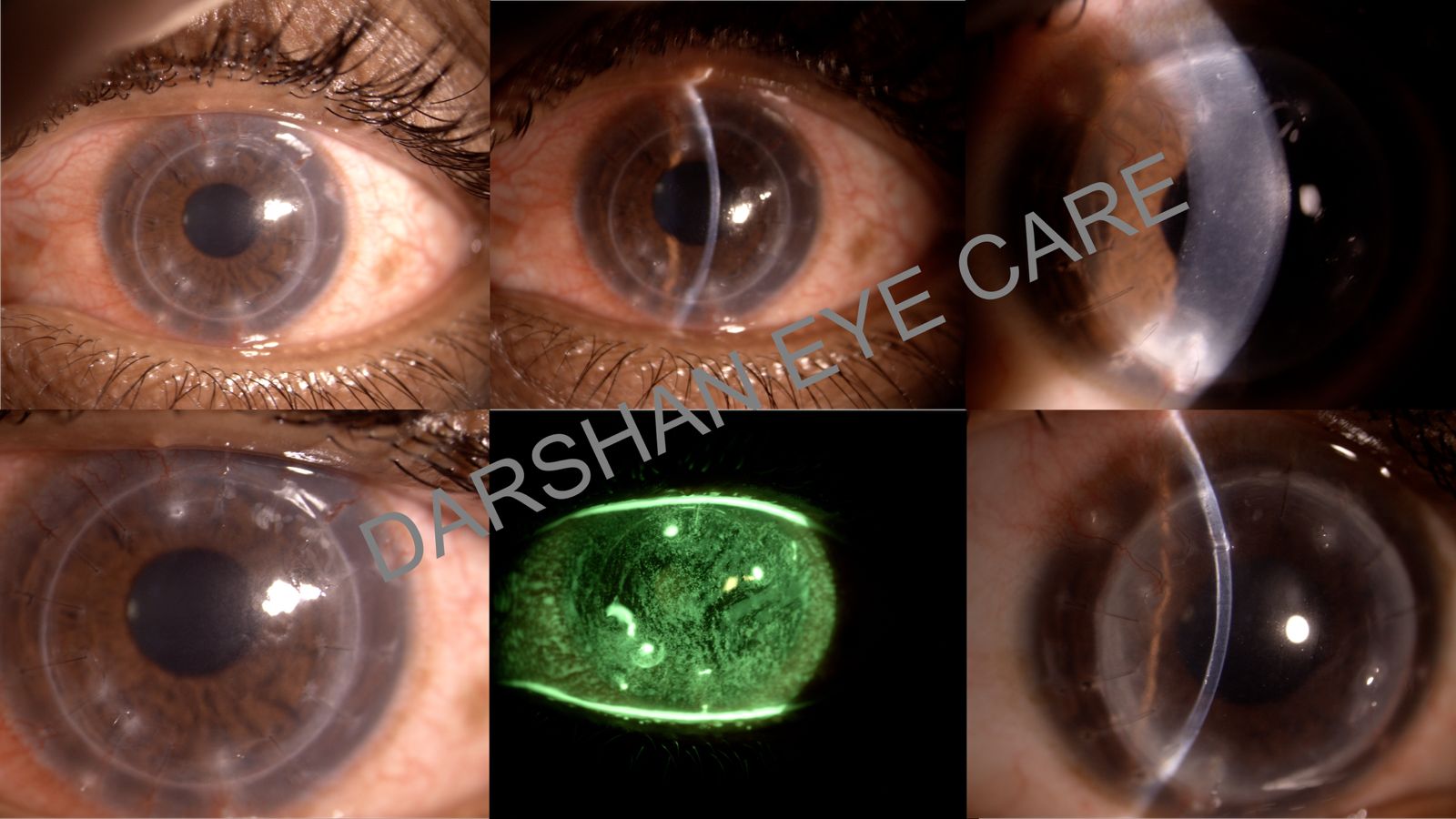
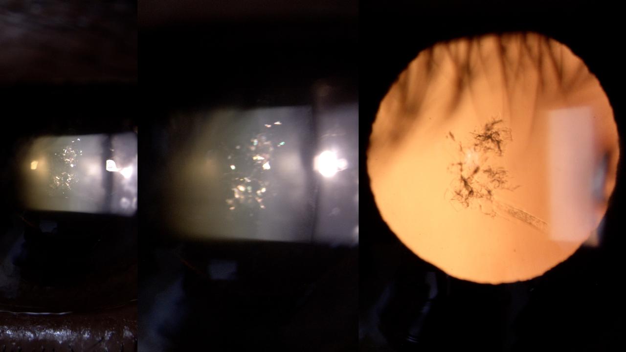
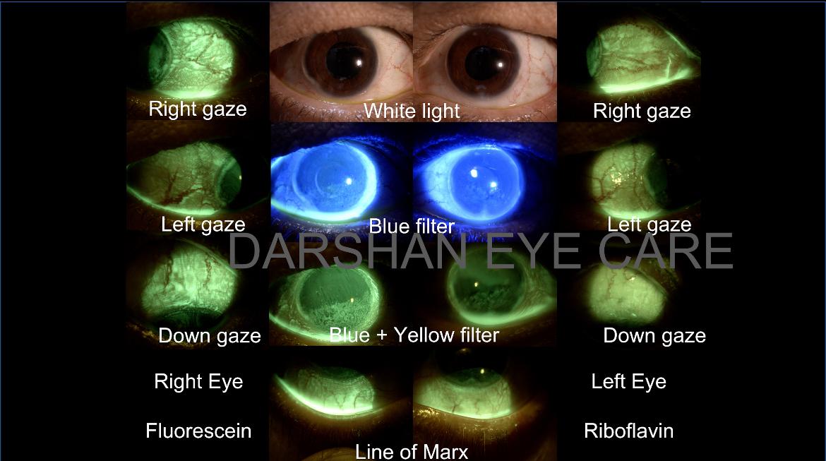
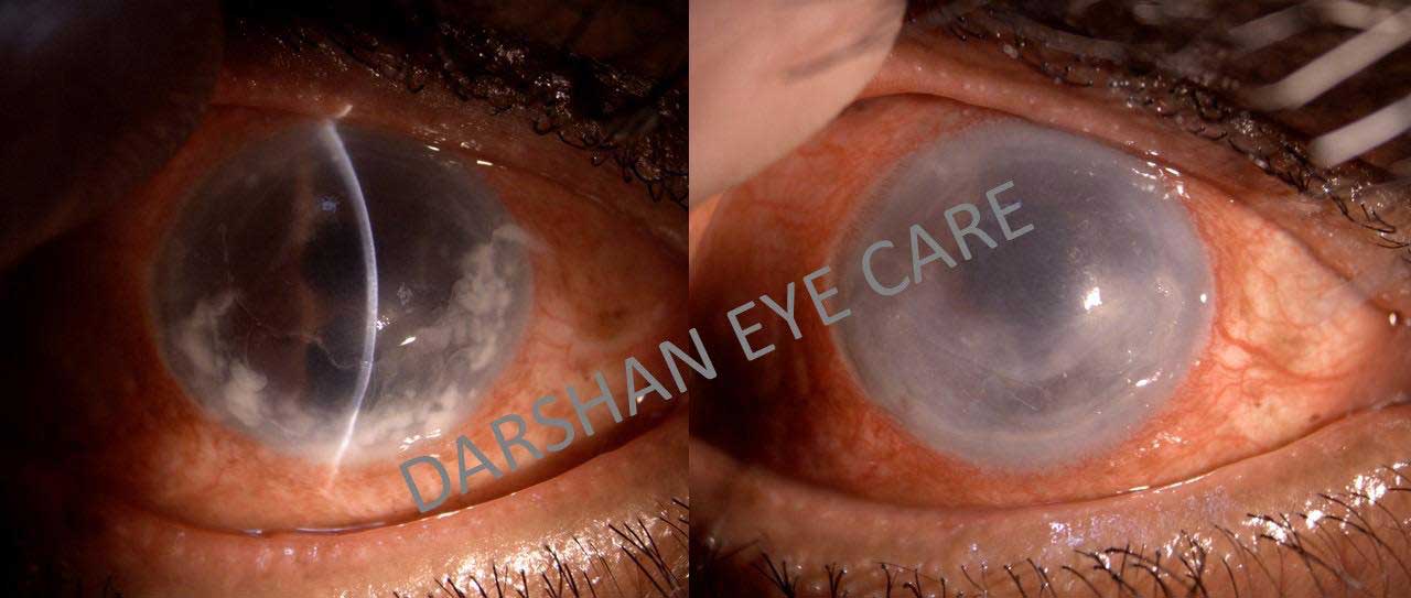
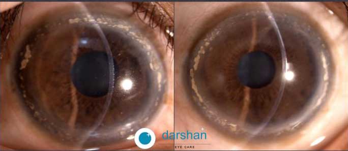
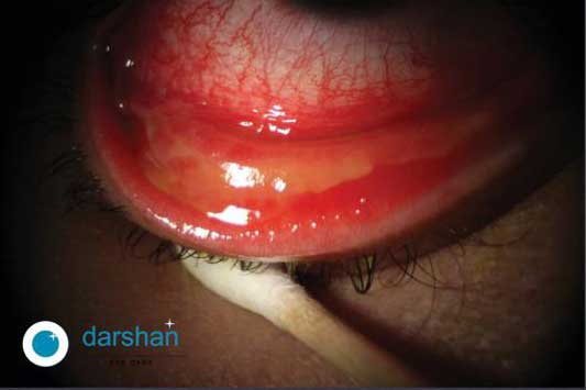
Conjunctival Melanosis Clinical Challenges Pigmented lesions of the conjunctiva are a common finding during an ophthalmic examination. They can pose a significant diagnostic challenge since they can stem from a wide variety of causes. Although malignant melanomas of the conjunctiva are rare, and account for less than 5% of ocular melanoma (majority are from the uveal tract), they can have significant morbidity and mortality and hence, early diagnosis is important. When evaluating these lesions, the clinical appearance, age at presentation, location, bilaterally and when in doubt, histopathology can help. In a 10-year-old boy, with dark skin, brown irides, the appearance of light brown, dispersed flat lesions seen symmetrically in both eyes, with no change since detection, the diagnosis is Complexion associated Melanosis (formerly called racial melanosis). Documentation and periodic observation for change in size, color, thickness and vascularity are necessary.

DALK

DALK: Deep Anterior Lamellar Keratoplasty

0 Comments

  Clinical Challenges   Deep Anterior Lamellar Keratoplasty (DALK) Deep Anterior Lamellar Keratoplasty (DALK) retains the patients endothelium and hence is considered superior to a Penetrating Corneal transplant (PK) from the immunology point of view. However, patients undergoing DALK must also be advised to report early when symptoms of Redness, Sensitivity to light, Vision change or Pain (RSVP) occur. This patient who was 1 year post DALK, reported with redness for one month. Multiple loose and broken sutures are noted, with ocular surface epithelial changes. There is no infection, but early vascularization is noted in the deep stroma in the superior quadrant. Early suture removal, antibiotics to avoid infection, and steroids to reduce the inflammation and vascularization are required.

Christmas Tree Cataract

Christmas Tree Cataract – A Rare Ocular Finding

0 Comments

Clinical Challenges Christmas Tree Cataract – A Rare Ocular Finding A cataract is a common occurrence in an eye clinic. However, there are some unusual forms of lens opacification that are seen infrequently. One such type results in the formation of delicate, multi-colored crystals in the lens. It resembles a lighted christmas tree and is hence called a Christmas tree cataract. These changes result from an accumulation of cystine or cholesterol in the lens material. Causes include myotonic dystrophy, gout, diabetes with poor control, and increased calcium levels. Occasionally, it can also be an idiopathic age-related change and may affect only one eye. Recognition of this unusual type of lens change should alert the ophthalmologist to the presence of the aforementioned underlying causes. Relevant investigations are performed to look for and if present, treat the underlying conditions. When visually significant, the cataract is dealt with in the routine manner.

Riboflavin

Riboflavin staining of the Ocular Surface

0 Comments

Clinical Challenges Riboflavin staining of the Ocular Surface A new option is now available The ocular surface can be affected by a variety of problems. These can cause significant discomfort to the patient. One of the important ways in which the health of the ocular surface is assessed is by staining with dyes. Traditionally, fluorescein dye is used to assess the health of the cornea, while lissamine green and rose Bengal are used to study the health of the conjunctiva. In India, the dyes are available as strips, and sometimes, the latter two dyes do not elute from the strip well. All slit lamps have a blue filter and this is used to study the effects of fluorescein staining on the cornea. If fluorescein staining of the conjunctiva is to be assessed, an additional yellow filter must be used to enhance the staining. Recently, riboflavin has been used for staining of the ocular surface. Unlike fluorescein, it is available as a solution, which can be used from a bottle – with a shelf life of a month. With the blue and yellow filters, it can also be used to assess conjunctival staining.

Conjunctivitis

Severe Viral Conjunctivitis and Its Management

0 Comments

Severe Viral Conjunctivitis and Its Management   Clinical Challenges Although viral conjunctivitis is a common problem which occurs as epidemics, and often resolves without complications in 5 to 7 days,sometimes it can be more severe and result in complications. If it is very severe, either due to the strain of the virus involved, the immune reaction on the part of the patient’s eye, and or inappropriate medication, then it can result in the formation of conjunctival membranes. In such pseudomembranous conjunctivitis, there is a need for topical steroid therapy, removal of the membranes, and protection to the corneal surface with antibiotics. Unpreserved lubricants and cold compresses can also help. However, conjunctival shrinkage and late-onset dry eye are concerns if the above treatment is not instituted in time.

Fungal keratitis

Fungal keratitis

0 Comments

  Clinical Challenges   Fungal keratitis Fungal keratitis is not uncommon in India, and this is probably due to a combination of the hot tropical climate and a largely agrarian economy. Injury with vegetative matter is an important cause for fungal keratitis. Recognizing the fungal cause of the corneal infection is extremely important, as it does not respond to the commonly used first-line fourth generation fluoroquinolone antibiotic drops, unlike bacterial keratitis. A delay in recognizing the fungal cause of the keratitis, and instituting the appropriate antifungal agents, can result in significant corneal morbidity and vision loss. A study from Aravind Eye Care system, has shown that much of the fungal infections in South India, are due to filamentous organisms, and they respond better to natamycin eye drops, rather than voriconazole eye drops. These infections typically have a dry stromal infiltrate, with hyphate edges, and satellite lesions, and the signs can exceed symptoms at the start of the infection. Yeast infections, usually by candida, are rare and are seen in immunocompromised patients. These infections can be multiple, have fluffy stromal infiltrates and do not respond to natamycin eye drops. They respond better to fluconazole, amphotericin B and voriconazole eye drops. Hence, determining the fungal cause of infectious keratitis, and the type of fungal organism at an early stage helps institute appropriate therapy and promotes corneal healing. This will require corneal scrapings and cultures. Darshan Google Reviews

clinical challenges

Central Serous Chorioretinopathy (CSCR)

0 Comments

  Clinical Challenges   Central Serous Chorioretinopathy (CSCR) Central serous chorioretinopathy is an eye disease which often results in temporary visual impairment and is characterised by leakage of fluid under the retina, predominantly in the central macula. It is brough about by stress, smoking, steroid use, sleep disturbances and even natural events like pregnancy. If diagnosed early, it is easily treatable via Medications and lifestyle modifications. Incase, it is neglected, long term pooling of fluid in the macula can cause the death of nerve cells in the retina, resulting in permanent vision loss. In few patients, the condition may be recurrent and can occur either in the fellow or same eye.

Clinical Challenges

Uveitis – Causes, Symptoms & Advanced Treatments

0 Comments

  Clinical Challenges   UVEITIS Uveitis is a condition resulting from the inflammation of the blood vessels of the eye. It can be associated with conditions like autoimmune diseases, systemic infections, vascular disease or in some cases may be idiopathic (no discernible cause). Symptoms include pain, redness, sensitivity to light and diminution of vision. The treatment of uveitis requires a holistic approach which combines ocular examination, systemic evaluation and referral to specialities like rheumatology/immunology. Diagnosis requires a high index of suspicion, by those trained in this speciality. The treatment of the eye is usually concurrent with management of the systemic pathology and this ensures complete remission/cure.

Corneal Degenerations

0 Comments

  Clinical Challenges   Corneal Degenerations Terrien’s marginal degeneration is a peripheral corneal thinning disorder which results in loss of corneal shape and poor vision over time. It is typified by the presence of lipid deposits in the periphery and corneal vascularisation.Occasionally, it can be associated with symptoms of redness, pain and inflammation – as in this 35 year old lady. There is no known effective treatment for this condition and the importance of recognising the diagnosis is to help avoid unnecessary investigations and potentially dangerous treatment side effects.

Acute Conjuctivitis (Pink/madras Eye)

0 Comments

Clinical Challenges Acute Conjuctivitis (Pink/madras Eye) With The Onset Of Monsoon Season In Different Parts Of India, The Prevalence Of Acute Viral Conjuctivitis Is On The Rise. The Disease May Vary Between Mild Forms Which Settle Without Medications, To Severe Variants, Which May Require Treatment For Upto 3 Weeks Or More. It Can Affect Patients Of All Age Groups. Patients With Symptoms Of Red Eye, Itchiness And Discharge Are Advised To Visit The Hospital For A Consult And Are Advised Against The Use Of Over The Counter Eye Drops (Especially Those Containing Steroids) And Native Remedies, As The May Inadvertently Cause The Disease To Become Severe,thereby Prolonging Recovery.